Cervical Vertebrae Anatomy Diagram
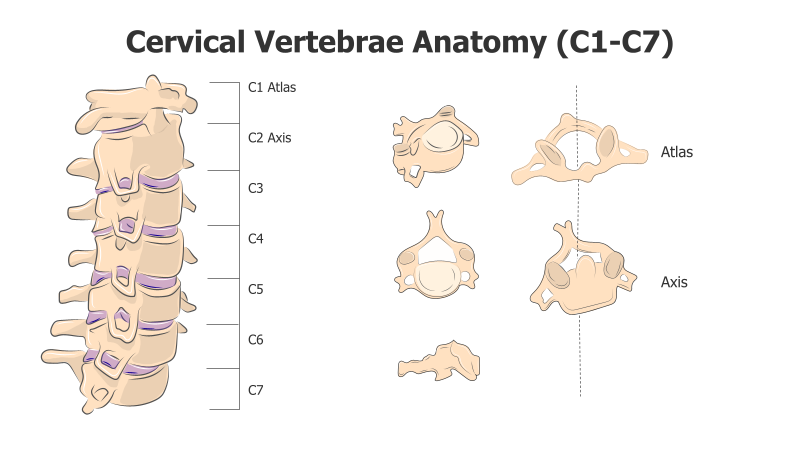
This template visually represents the anatomy of the cervical vertebrae (C1-C7). It showcases both a lateral view of the stacked vertebrae and individual cross-sections of key vertebrae.
Layout & Structure:
- The template features a stacked arrangement of seven vertebral levels (C1-C7) on the left, illustrating their position in the spinal column.
- To the right, individual cross-sections of the Atlas (C1) and Axis (C2) are displayed, alongside cross-sections of C3, C4, C5, C6, and C7.
- A vertical dashed line visually separates the stacked view from the individual cross-sections.
Style:
- The diagram employs a clean, illustrative style with a muted color palette.
- The vertebrae are depicted in a 3D-like rendering, providing a sense of depth and anatomical accuracy.
- The overall aesthetic is professional and informative, suitable for medical or educational presentations.
Use Cases:
- Medical education and training materials.
- Anatomy presentations for students and healthcare professionals.
- Patient education resources to explain spinal structure.
- Illustrating the differences between cervical vertebrae.
- Visual aids for chiropractic or physical therapy explanations.
Key Features:
- Clear depiction of cervical vertebrae anatomy.
- Highlights the unique features of the Atlas and Axis.
- Visually appealing and easy to understand.
- Suitable for a variety of educational and medical contexts.
Tags:
anatomycervical vertebraespinemedicaleducationdiagramskeletonatlasaxisc1c2vertebraehealthscience
Ready to Get Started?
Impress your audience and streamline your workflow with GraphiSlides!
Install Free Add-onNo credit card required for free plan.