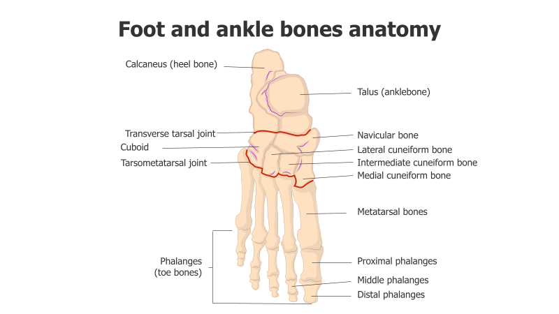
Foot and Ankle Bone Anatomy Diagram
This template presents a detailed anatomical diagram of the human foot and ankle, showcasing the various bones and joints.
Layout & Structure:
- The diagram features a skeletal representation of the foot and ankle, clearly labeling each bone.
- It includes the calcaneus (heel bone), talus (anklebone), navicular bone, cuboid, cuneiform bones (lateral, intermediate, and medial), metatarsal bones, and phalanges (proximal, middle, and distal).
- Key joints like the transverse tarsal joint and tarsometatarsal joint are also indicated.
- The arrangement is anatomical, providing a clear visual representation of bone placement.
Style:
- The diagram utilizes a clean, illustrative style with distinct bone outlines.
- Color-coding is used to differentiate bones and joints, enhancing clarity.
- Labels are concise and directly point to the corresponding anatomical structures.
- The overall aesthetic is professional and informative, suitable for educational or medical contexts.
Use Cases:
- Medical education and training materials.
- Patient education resources to explain foot and ankle anatomy.
- Orthopedic presentations and reports.
- Anatomy textbooks and reference materials.
- Podiatry consultations and treatment planning.
Key Features:
- Accurate anatomical representation.
- Clear and concise labeling.
- Visually appealing and easy to understand.
- Suitable for both print and digital presentations.
- Provides a comprehensive overview of foot and ankle bone structure.
Tags:
anatomyfootanklebonesskeletonmedicaldiagramillustrationorthopedicspodiatryjointscalcaneustalusphalangesmetatarsalscuneiform
Ready to Get Started?
Impress your audience and streamline your workflow with GraphiSlides!
Install Free Add-onNo credit card required for free plan.