Thyroid-Parathyroid Gland Anatomy
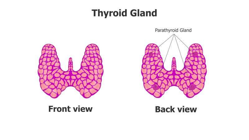
This slide visually represents the anatomy of the thyroid and parathyroid glands, showcasing both front and back views.
Layout & Structure:
- The slide features two diagrams: a front view and a back view of the thyroid gland.
- The thyroid gland is depicted as a bilobed structure, connected by an isthmus.
- The parathyroid glands are shown as smaller structures located on the posterior surface of the thyroid gland.
- The diagrams are clearly labeled with "Front view" and "Back view" for easy understanding.
Style:
- The diagrams utilize a consistent color scheme (purple/pink) and a textured fill to represent the glandular structure.
- The style is clean and illustrative, suitable for educational or medical presentations.
- The use of labels and arrows enhances clarity and directs the viewer's attention.
Use Cases:
- Medical education and training materials.
- Presentations on endocrine system anatomy.
- Patient education resources.
- Anatomy and physiology lectures.
- Visual aids for healthcare professionals.
Key Features:
- Clear and accurate anatomical representation.
- Easy-to-understand labeling.
- Visually appealing and informative design.
- Suitable for a variety of presentation formats.
Tags:
thyroidparathyroidanatomyglandendocrinemedicalbiologyhealthdiagramfront viewback view
Ready to Get Started?
Impress your audience and streamline your workflow with GraphiSlides!
Install Free Add-onNo credit card required for free plan.