Pancreas Anatomy Illustration
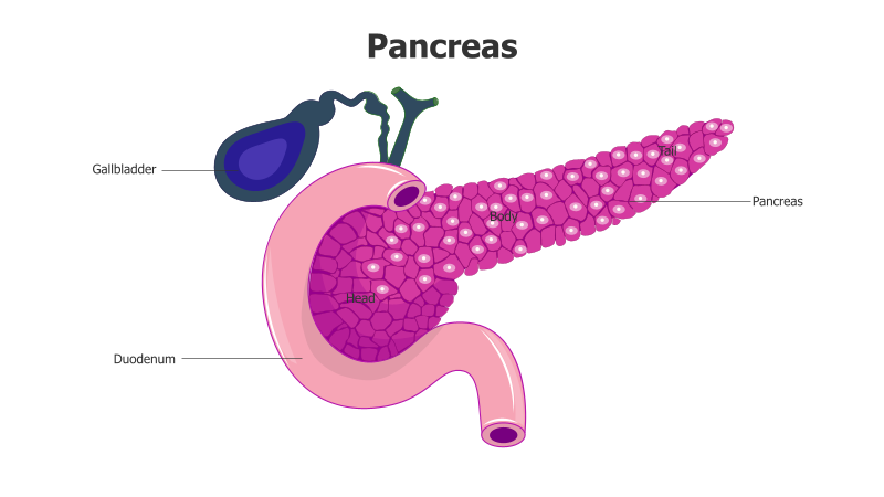
This slide presents a detailed anatomical illustration of the human pancreas and surrounding organs.
Layout & Structure: The slide features a diagram of the pancreas, clearly labeled with its three main parts: head, body, and tail. It also shows the gallbladder and duodenum, illustrating their relationship to the pancreas. The pancreas is depicted in a detailed, textured style, with internal structures visible. The overall layout is straightforward and focuses on anatomical accuracy.
Style: The illustration employs a realistic, medical illustration style with a color palette of pink, purple, and green. The use of shading and texture gives the diagram a three-dimensional appearance. The style is professional and informative, suitable for educational or medical presentations.
Use Cases:
- Medical education and training
- Presentations on digestive system anatomy
- Patient education materials
- Scientific publications and research
- Health and wellness presentations
Key Features:
- Clear labeling of key anatomical structures
- Detailed and accurate illustration
- Visually appealing and informative design
- Suitable for a variety of medical and educational contexts
- Illustrates the relationship between the pancreas, gallbladder, and duodenum
Tags:
Ready to Get Started?
Impress your audience and streamline your workflow with GraphiSlides!
Install Free Add-onNo credit card required for free plan.