Kidney Anatomy Detailed Diagram
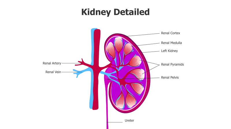
This slide presents a detailed anatomical diagram of the human kidney.
Layout & Structure:
- The slide features a cross-sectional view of the left kidney, showcasing its internal structures.
- Key components are clearly labeled with connecting lines, including the renal cortex, renal medulla, renal pyramids, renal pelvis, ureter, renal artery, and renal vein.
- The kidney is depicted in a semi-circular shape with internal structures arranged to illustrate their spatial relationships.
Style:
- The diagram utilizes a vibrant color palette with shades of pink, purple, red, and blue to differentiate structures.
- A clean, illustrative style is employed, providing a clear and understandable representation of kidney anatomy.
- The overall aesthetic is professional and educational.
Use Cases:
- Medical education and training materials.
- Presentations on the urinary system and kidney function.
- Patient education resources.
- Anatomy and physiology lectures.
- Healthcare presentations and reports.
Key Features:
- Detailed and accurate anatomical representation.
- Clear labeling of key kidney structures.
- Visually appealing and easy-to-understand diagram.
- Suitable for a variety of educational and medical contexts.
Tags:
kidneyanatomymedicalbiologydiagramrenalureterarteryveinhealthscienceeducation
Ready to Get Started?
Impress your audience and streamline your workflow with GraphiSlides!
Install Free Add-onNo credit card required for free plan.