Human Lungs Anatomy
This slide illustrates the anatomy of the human lungs, showcasing their lobes and associated structures.
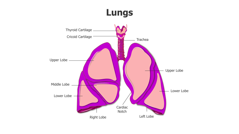
Layout & Structure: The slide features a detailed illustration of the lungs, trachea, and associated cartilages. The lungs are depicted with their major lobes clearly labeled: upper, middle (right lung only), and lower lobes. The cardiac notch is also indicated. The trachea and thyroid/cricoid cartilages are shown at the top, providing context for the respiratory system's upper airways.
Style: The illustration employs a clean, modern aesthetic with a color scheme of purple and pink. The use of solid colors and clear labeling creates a professional and informative visual. The depiction is realistic but simplified for clarity.
Use Cases:
- Medical education and presentations
- Anatomy and physiology lessons
- Healthcare infographics
- Patient education materials
- Biology and health science courses
Key Features:
- Clear and accurate anatomical depiction
- Well-defined labels for easy understanding
- Visually appealing color scheme
- Suitable for a wide range of educational contexts
- Illustrates key components of the respiratory system
Tags:
Ready to Get Started?
Impress your audience and streamline your workflow with GraphiSlides!
Install Free Add-onNo credit card required for free plan.