Liver Anatomy Diagram
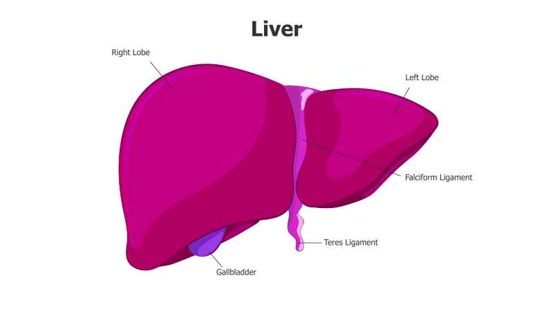
This slide presents a detailed anatomical diagram of the human liver.
Layout & Structure: The diagram showcases the liver's primary lobes – the right and left lobes – along with associated structures like the gallbladder, falciform ligament, and teres ligament. The liver is depicted as a large, irregularly shaped organ, with the gallbladder positioned inferiorly. Ligaments are shown as connecting structures.
Style: The diagram employs a flat, illustrative style with solid color fills. The use of a vibrant pink hue for the liver and a contrasting purple for the gallbladder and ligaments creates visual distinction. The labels are clear and concise, enhancing readability. The overall aesthetic is clean and informative.
Use Cases:
- Medical education and presentations
- Anatomy and physiology lessons
- Healthcare professional training
- Patient education materials
- Illustrating liver-related conditions
Key Features:
- Clearly labeled anatomical structures
- Visually appealing and informative design
- Suitable for a wide range of educational purposes
- Easy to understand and interpret
Tags:
Ready to Get Started?
Impress your audience and streamline your workflow with GraphiSlides!
Install Free Add-onNo credit card required for free plan.