Human Heart Anatomy
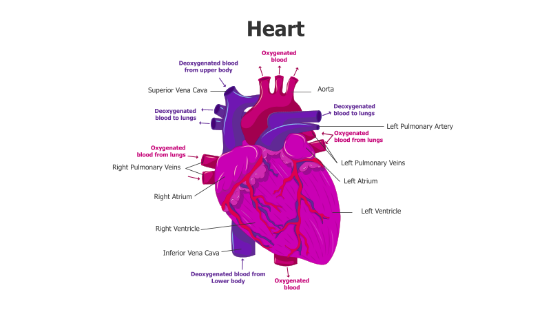
This slide illustrates the anatomy of the human heart, showcasing its chambers, major blood vessels, and blood flow pathways.
Layout & Structure: The slide features a detailed, labeled diagram of the heart. Key components like the right and left atria, right and left ventricles, aorta, vena cava (superior and inferior), and pulmonary arteries/veins are clearly identified. Arrows indicate the direction of blood flow, distinguishing between oxygenated and deoxygenated blood. The heart is centrally positioned with labels radiating outwards.
Style: The diagram employs a realistic, illustrative style with color-coding to differentiate oxygenated (red) and deoxygenated (blue) blood. The use of shading and detail provides a visually engaging and informative representation of the heart's structure. The overall aesthetic is professional and educational.
Use Cases:
- Medical education and presentations
- Biology lessons and materials
- Health and wellness content
- Patient education materials
- Anatomy and physiology studies
Key Features:
- Clear and accurate anatomical representation
- Color-coded blood flow pathways
- Detailed labeling of key heart components
- Visually engaging and informative
- Suitable for a wide range of educational purposes
Tags:
Ready to Get Started?
Impress your audience and streamline your workflow with GraphiSlides!
Install Free Add-onNo credit card required for free plan.